

Introduction of academicians

Teng Gaojun
Interventional radiologists and radiologists
The only academician of the Chinese Academy of Sciences in the field of interventional radiology, Teng Gaojun, is currently the president of Zhongda Hospital affiliated to Southeast University and the director of the Interventional Diagnosis and Treatment Center. He is also the director of the Key Laboratory of Molecular Imaging and Functional Imaging in Jiangsu Province and the president of the Interventional Physicians Branch of the Chinese Medical Association. Professor Teng has pioneered more than 10 new interventional technologies and completed tens of thousands of interventional surgeries. He has also made significant contributions to international interventional radiology and became the first Chinese person to receive the Gold Award from the American Society of Interventional Radiology, the highest honor in the field of interventional radiology awarded by the society. In addition, he has also been awarded the highest honor awards by the European Society of Cardiovascular Interventional Radiology and the Asia-Pacific Society of Cardiovascular and Interventional Radiology.
On November 14, 2022, the team led by academician Teng Gaojun used the cutting-edge irreversible electroporation ablation technology to eliminate tumors for patients. During the surgery, they used the "new domestic Nanoknife" - the High Voltage Steep Pulse Minimally Invasive Therapeutic System independently developed and produced by Shanghai Yuanshan Medical Technology Co., Ltd. (hereinafter referred to as "Alpmed").
The patient, an 83-year-old male, was admitted with complaints of loss of appetite, yellowing of urine, and aversion to oily food for over a month. MRI examination at an external hospital showed abnormal signals in the right lobe of the liver, as well as gallstones in the right lobe of the liver, gallbladder, and common bile duct. MRI cholangiopancreatography suggested cholangiocarcinoma. PET-CT showed a significant increase in SUV of the lesion and elevated tumor marker CA199, and a biopsy was considered for cholangiocarcinoma.
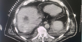
(Preoperative imaging)
After consultation with the patient, Academician Teng Gaojun and his team stated that the patient is of advanced age and has a complex medical condition, including atrial fibrillation with second-degree atrioventricular block, hypertension, coronary heart disease, and severe stenosis in the P2 segment of the right posterior cerebral artery. The tumor is located close to the hepatic vein and portal vein, making conventional tumor resection or microwave ablation surgery risky or difficult to completely ablate. NanoKnife tumor ablation therapy is a new and advanced technology in tumor ablation, which differs from traditional thermal ablation techniques in its mechanism of inactivation. It can selectively ablate tissues without being affected by thermal sink effects, effectively preserving important tissue structures such as blood vessels, nerves, bile ducts, and ureters, resulting in more thorough and precise tumor inactivation.

Surgery Introduction
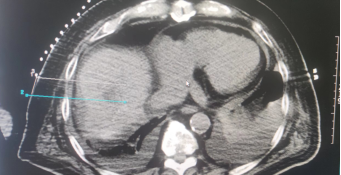
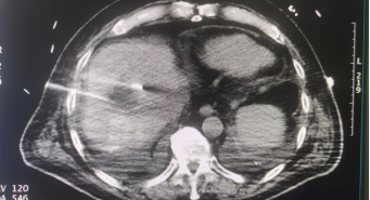
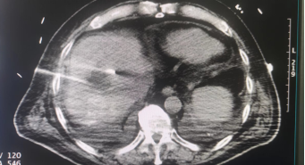
On November 14th, Academician Teng Gaojun led and guided his team to perform NanoKnife tumor ablation surgery on the patient. The patient was in a supine position and received needle puncture. The tumor had a maximum diameter of 3cm, with a 2cm interval between each parallel tumor. The needle tip of the ablation needle was exposed by 1.5cm to ensure complete coverage of the ablation area around the tumor before starting the ablation. During the ablation process, the equipment energy field was stable, the voltage almost did not attenuate, and the current steadily increased. The ablation was completed smoothly in more than ten minutes. Finally, the needle was withdrawn, and a CT enhanced scan was performed. The tumor showed low density with visible gasification around it, indicating a successful surgery. The patient recovered and was discharged on November 25th.
(Intraoperative needle placement diagram)

(Immediate postoperative imaging)
Academician Teng Gaojun stated that irreversible electroporation (IRE) technology is the most advanced treatment method for tumor ablation. This technology not only provides a new treatment option for patients with cancer in special locations (such as the hepatic hilum and pancreatic head) who cannot undergo traditional surgical methods (such as resection and thermal ablation), but also studies have shown that IRE technology can be combined with immunotherapy and radiotherapy/chemotherapy to further improve its therapeutic effect. This not only adds new treatment methods to the department, but also benefits more cancer patients by providing them with new treatment options and new hope.

 Membership